
What is the difference between C-TAME and s-TAME? A complete comparison of catheter-based and ultrasound-guided transcatheter arterial microembolization
What is the difference between C-TAME and s-TAME? A complete comparison of catheter-based and ultrasound-guided transcatheter arterial microembolization
Article Summary
Both C-TAME and s-TAME belong to TAME (microvascular embolization therapy) and share the same therapeutic purpose, which is to treat potential abnormal microvessels at chronic pain sites. The main difference lies in the treatment methods and guidance tools: C-TAME is performed via a catheter combined with angiography equipment, making it suitable for deeper areas or those requiring precise localization; s-TAME uses ultrasound-guided fine-needle injections, making it suitable for superficial pain areas that can be localized by ultrasound. They are not old and new versions of each other; rather, the choice of method is based on the location, depth of pain, and assessment results.
Chronic pain that has recurred for more than three months with limited response to other treatments is most suitable for evaluation.
What is TAME?
TAME (Transcatheter Arterial Micro-Embolization) is a minimally invasive treatment targeting the mechanisms of chronic pain. When pain persists for a long time, abnormal proliferation of microvessels and associated nerve changes may appear at the site of pain, contributing to the formation and maintenance of chronic pain. The approach of TAME is to selectively treat pain-related abnormal microvessels using embolic materials to reduce the influence of these vascular-related mechanisms on pain, rather than simply suppressing the sensation of pain.
The TAME technology was developed and promoted by Dr. Yuji Okuno in Japan, and related studies have been published in international medical journals. Hee Clinic has obtained the Taiwan trademark authorization from Okuno Clinic in Japan to provide evaluations for catheter-based C-TAME and ultrasound-guided s-TAME based on different pain locations and medical conditions.
What is the difference between C-TAME and s-TAME? Understand it in one table
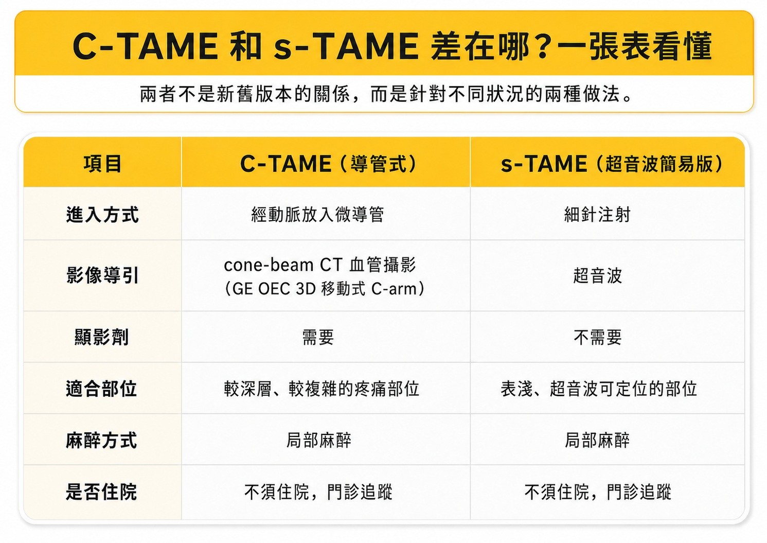
C-TAME accesses the target area via a catheter and uses angiography to confirm the location, making it suitable for treating deeper areas. s-TAME uses a fine needle guided by ultrasound, which makes the procedure simpler, but it is only suitable for superficial areas that can be seen under ultrasound. Both are outpatient procedures under local anesthesia and do not require hospitalization.
The difference between the two is not about "which one is better," but rather about the areas they are suitable for treating and the required conditions. The catheter in C-TAME can reach deep areas and confirm the embolization range under angiography; s-TAME eliminates catheters and contrast agents, making the process simpler, but it is only applicable to superficial targets visible on ultrasound.
Why does C-TAME require angiography equipment?
C-TAME requires angiography equipment to assist physicians in locating target blood vessels.
Since the microvessels associated with chronic pain are small in diameter, it may be difficult to confirm the catheter position and embolization range relying solely on flat images. Angiography equipment equipped with cone-beam CT functions can provide 3D imaging information during the treatment process, helping physicians locate blood vessels more accurately for selective embolization.
Hee Clinic is equipped with the GE OEC 3D mobile C-arm with cone-beam CT functionality to perform C-TAME evaluation and treatment processes in our in-house angiography operating room, eliminating the need for patients to be referred to other institutions for related procedures.
In contrast, s-TAME uses ultrasound as a guidance tool and does not require angiography equipment, so it is mainly applied to superficial pain areas that can be clearly localized by ultrasound.
Which situations are suitable for evaluating C-TAME? Which are suitable for s-TAME?
The choice between C-TAME and s-TAME mainly depends on the location and depth of pain, as well as whether the relevant blood vessels can be precisely located and treated.
If the pain is located in deeper areas or requires angiography to confirm the target blood vessels, the physician may evaluate whether C-TAME is suitable. Examples include: chronic knee pain caused by degenerative knee osteoarthritis, frozen shoulder (adhesive capsulitis), chronic pain around the hip joint, and recurrent pain around deep joints.
If the pain location is superficial and can be clearly localized via ultrasound, it may be suitable to evaluate s-TAME. Examples include: De Quervain's tenosynovitis (mommy's wrist), plantar fasciitis, tennis elbow, finger osteoarthritis, and chronic pain at tendon insertions.
The actual suitable method must still be determined by a physician after consultation, physical examination, and imaging evaluation. For some patients, the evaluation may reveal that the cause of pain does not stem from a mechanism suitable for embolization, and other treatment directions may be recommended.
Can s-TAME also be done on the knee? Under what circumstances would the catheter method be recommended?
An evaluation can be performed.
What decides the treatment method is not the "knee" area itself, nor which type of clinic it is performed in. The key lies in three things: whether the pain source matches abnormal blood flow, the depth and branching status of the target vessels, and whether the imaging can clearly show the needle or catheter path.
The accumulation of literature for the two approaches is not the same. Direct puncture with ultrasound-guided fine needles currently has a more complete clinical follow-up series focused on superficial areas such as plantar fasciitis. Embolization studies for the knee joint are primarily guided by angiography, often referred to in international literature as GAE (Genicular Artery Embolization), which includes randomized trials with sham controls—some showing supportive results and others showing no significant difference. The reason is easy to understand: the genicular arteries have many branches and complex anastomoses, so most formal studies choose methods that can visualize the complete vascular map.
Hee Clinic performs both, and there is no need to lean toward either side during evaluation. For targets that are superficial, clearly identifiable by ultrasound, and have a safe needle insertion path, s-TAME is often among the options. For cases that are deeper, have more branches, and require real-time angiography or 3D imaging for confirmation, the catheter method will be recommended. The catheter-based C-TAME performed on the knee joint at Hee Clinic is likewise conducted under angiography guidance.
There is also a third answer: do neither for the time being.
If the source of pain does not match the abnormal blood flow, performing any kind of embolization is just doing an extra treatment. Finding out the exact cause always comes before choosing a treatment method.
Treatment Process and Recovery of C-TAME and s-TAME
In most cases, C-TAME is performed under local anesthesia. The physician will establish vascular access from the wrist or another suitable location, deliver the microcatheter to the target vessel, and then perform selective embolization. The treatment wound is usually only the size of a pinhole. Patients can return home after observation, and follow-up is arranged based on the treated area and individual conditions.
Under ultrasound guidance, s-TAME uses a fine needle to localize and treat the pain-related area. Since there is no need to establish vascular access, the overall process is relatively simple, but its applicability depends on whether the target area can be clearly localized by ultrasound. The actual treatment method and recovery time must still be explained by the physician according to individual conditions.
Frequently Asked Questions
Q: Is C-TAME a surgery?
C-TAME is not a traditional open surgery, but a minimally invasive interventional therapy performed through blood vessels. The treatment establishes access through an arterial pinhole without the need to cut tissue, and is performed under local anesthesia in most cases.
Q: Will the effect of s-TAME be worse than C-TAME?
C-TAME and s-TAME deal with different target conditions and are not an upgraded version of one another. If the pain location is suitable for ultrasound localization, s-TAME may be the right choice; if it is necessary to enter deeper vessels or locate them more precisely, C-TAME may be required.
Q: Can I still undergo TAME if I have had PRP or hyaluronic acid injections?
An evaluation can be performed. Having received previous injection treatments does not affect your eligibility for a TAME evaluation. The physician will make a judgment based on your current condition and imaging.
Q: How many times does TAME need to be done?
The number of treatments varies depending on the pain location, medical condition, and response to treatment. It cannot be generalized and must be explained by the physician after evaluation.
Q: Can s-TAME be done for knee degeneration?
An evaluation can be performed, but it is not suitable for everyone. The criteria for judgment are the source of pain, the depth of the target vessels, and imaging conditions, not the area itself. For vascular targets that are superficial and clearly identifiable by ultrasound, s-TAME can be considered; when the location is deep or the vascular branches are complex, the catheter-based method under angiography guidance is mostly recommended. Hee Clinic performs both methods, which will be discussed with you after a physician's evaluation. It is also possible that neither method will be recommended after evaluation.
Q: Where in Taipei can I get evaluated for C-TAME?
Hee Clinic provides evaluation services for both C-TAME and s-TAME. Whether you are suitable for treatment must be determined by a physician after consultation, physical examination, and imaging evaluation.
Hee Clinic is located at 2F-1, No. 40, Sec. 2, Heping E. Rd., Da'an Dist., Taipei City. It operates entirely on an appointment basis. If you require a chronic pain evaluation, you can schedule an appointment for a pain clinic session via LINE or by calling (02) 2366-0186.
Yuji Okuno Microvascular Embolization References
Okuno Y, et al. Midterm clinical outcomes and MR imaging changes after transcatheter arterial embolization as a treatment for mild to moderate radiographic knee osteoarthritis resistant to conservative treatment. J Vasc Interv Radiol. 2017;28(7):995-1002. doi:10.1016/j.jvir.2017.02.033
Written by: Dr. Ping-Sheng Lu
Last updated: 2026/08/10
